Anatomy and physiology lab manuals provide interactive, applied learning experiences, such as the 7th Edition, offering step-by-step guides and experiments to help students grasp complex biological concepts effectively․
1․1 Importance of Lab Work in Anatomy and Physiology
Lab work is essential in anatomy and physiology as it provides hands-on experience, reinforcing theoretical knowledge through practical experiments․ It enhances understanding of complex biological structures and processes, such as tissue identification and organ function․ Interactive learning fosters critical thinking and problem-solving skills, preparing students for real-world applications in healthcare and research․
1․2 Safety Protocols in the Lab
Safety protocols are crucial in anatomy and physiology labs to protect students and equipment․ These include wearing protective gear like gloves and goggles, proper handling of biological specimens, and adherence to disposal guidelines․ Emergency procedures, such as spill management and fire evacuation plans, are also essential to ensure a secure learning environment․
Anatomical Terminology
Anatomical terminology provides a universal language for describing body structures and their locations, ensuring clear communication in scientific and medical contexts, as outlined in lab manuals․
2․1 Directional Terms and Planes of the Body
Directional terms like anterior, posterior, medial, and lateral provide a standardized language to describe body structures․ The three planes of the body—sagittal, frontal, and transverse—help in visualizing and sectioning the body․ These terms and planes are essential for accurate communication in anatomy, aiding in dissection and identification of structures during lab work, as detailed in anatomy and physiology lab manuals․
2․2 Understanding Anatomical Positions
The standard anatomical position is a reference point where the body stands upright, facing forward, with arms at the sides and palms facing forward․ This position helps maintain consistency in describing body structures․ Lab manuals emphasize understanding anatomical positions to accurately identify and describe locations of organs and tissues during dissection and study, ensuring clear communication among students and professionals․
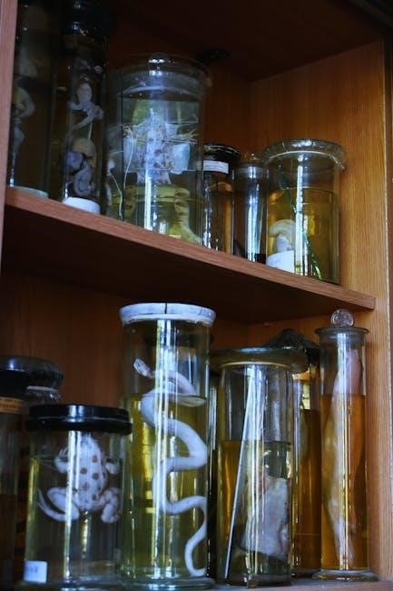
Histology and Microscopy
Histology involves studying tissues under microscopes, a crucial lab skill in anatomy and physiology․ Microscopy helps examine cellular structures, essential for understanding tissue functions and diagnoses․
Microscopes are essential tools in anatomy and physiology labs, enabling detailed examination of cellular structures․ Compound and stereomicroscopes are commonly used for studying tissue samples․ Proper handling and focusing techniques are critical for clear observations․ Histology labs often involve preparing slides and using microscopes to identify cellular details, making them indispensable for understanding tissue functions and diagnostic processes․
3․2 Preparing Tissue Samples for Examination
Preparing tissue samples involves fixing, sectioning, and staining to ensure clear microscopic observation․ Fixing preserves tissue structure, while sectioning creates thin slices using a microtome․ Staining enhances contrast, revealing cellular details․ Proper preparation is critical for accurate histological analysis, ensuring that samples are suitable for study under a microscope․
Skeletal System
The skeletal system consists of 206 bones, providing support, protection, and movement․ Lab manuals explore bone structure, types, and functions through experiments and dissection activities․
4․1 Structure and Function of Bones
Bones are rigid, calcified tissues forming the skeleton, providing structural support and protection․ They consist of cortical (compact) and trabecular (spongy) bone, with marrow producing blood cells․ Their functions include movement, mineral storage, and blood cell production․ Lab manuals often include bone dissection and histology studies to explore their microscopic structure and classify them into long, short, flat, and irregular types;
4․2 Classification of Joints and Their Movements
Joints are points where bones connect, enabling movement․ They are classified into fibrous, cartilaginous, and synovial joints․ Synovial joints, the most mobile, include hinge, ball-and-socket, pivot, and gliding types․ Lab manuals often include activities to identify joint types and their ranges of motion, such as flexion, extension, rotation, and circumduction, using diagrams and models to enhance understanding of joint functionality and mobility․
Muscular System
The muscular system consists of three types of muscles: skeletal, smooth, and cardiac․ Lab manuals explore their structure, functions, and roles in movement, support, and bodily processes․
5․1 Types of Muscles and Their Functions
The muscular system comprises three types of muscles: skeletal, smooth, and cardiac․ Skeletal muscles, attached to bones, enable voluntary movements․ Smooth muscles, found in internal organs, facilitate involuntary functions like digestion․ Cardiac muscles, exclusive to the heart, ensure continuous blood circulation․ Lab manuals detail their unique structures and roles, providing hands-on activities to explore their functions and significance in human physiology․
5․2 Physiological Responses of Muscles
Muscles respond to stimuli through contraction, relaxation, and rhythmic movements․ Skeletal muscles react to nerve impulses, enabling voluntary actions like walking․ Smooth muscles function involuntarily, regulating processes such as digestion․ Cardiac muscles sustain heartbeat rhythms․ Lab manuals explore these physiological responses, offering exercises to study muscle contractions, nerve stimulation, and energy sources like ATP, enhancing understanding of muscle dynamics and their vital roles in movement and bodily functions․
Nervous System
The nervous system consists of the brain, spinal cord, and nerves, controlling voluntary and involuntary body functions․ Lab manuals explore nerve responses and neural communication through experiments․
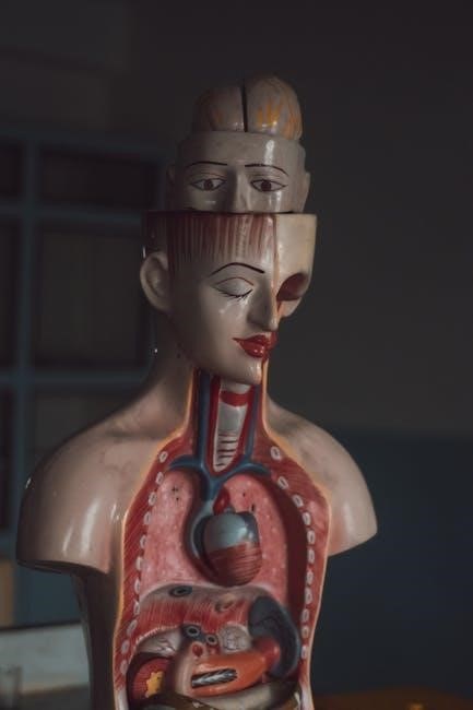
6․1 Structures and Functions of the Brain
The brain, the control center of the body, comprises the cerebrum, cerebellum, and brainstem․ Lab manuals include activities like brain dissection, neural mapping, and case studies to explore cognitive functions, motor control, and sensory processing․ These exercises help students understand the brain’s role in regulating body systems and its complex neural pathways․
6․2 Roles and Functions of the Spinal Cord
The spinal cord acts as a communication pathway between the brain and body, facilitating reflexes and controlling involuntary functions like heart rate and digestion․ Lab manuals often include dissection exercises and histological studies to explore its structure․ Activities focus on understanding its role in motor and sensory functions, as well as its clinical relevance in injuries affecting movement and sensation․
Digestive System
The digestive system, explored in lab manuals, involves key organs like the stomach, intestines, and liver, focusing on processes like digestion, absorption, and nutrient utilization through practical experiments․
7․1 Key Organs and Their Roles
The digestive system comprises essential organs, each with unique functions․ The mouth initiates digestion with teeth and enzymes, while the esophagus transports food to the stomach․ The stomach churns food with gastric juices, breaking it down further․ The small intestine absorbs nutrients into the bloodstream, supported by bile from the liver and enzymes from the pancreas․ The large intestine absorbs water, and waste is eliminated through the rectum․
7․2 Processes of Digestion and Absorption
Digestion begins with mechanical breakdown in the mouth and chemical digestion via enzymes like amylase․ Gastric juices in the stomach further break down food into a liquid mixture․ In the small intestine, enzymes from the pancreas and bile from the liver facilitate nutrient absorption․ Villi increase surface area for efficient absorption․ The large intestine absorbs water, forming feces for elimination, completing the digestive process․
Clinical Applications in Anatomy and Physiology
Clinical applications bridge anatomy and physiology with real-world healthcare scenarios, enabling students to understand how lab concepts translate to patient care, diagnosis, and treatment․
8․1 Case Studies in Clinical Anatomy
Clinical anatomy case studies provide real-world examples, enabling students to analyze conditions like injuries or diseases; These scenarios help apply anatomical knowledge to diagnose and treat conditions, emphasizing clinical relevance and practical skills for future healthcare professionals, while enhancing critical thinking and problem-solving abilities through hands-on learning experiences․
8․2 Diagnostic Techniques and Procedures
Diagnostic techniques in anatomy and physiology involve imaging methods like X-rays, MRIs, and CT scans to visualize internal structures․ Procedures such as biopsies and endoscopies are also used to examine tissues and organs․ These tools are essential for accurate diagnoses, helping students understand how anatomical knowledge translates into clinical practice, enhancing their ability to identify and treat medical conditions effectively․
Dissection and Lab Techniques
Dissection and lab techniques are crucial for understanding anatomical structures․ Essential tools like scalpels, forceps, and magnifiers are used to explore tissues and organs systematically․
9․1 Essential Tools and Equipment for Dissection
Essential tools for dissection include scalpels, forceps, magnifiers, and dissection trays․ Protective gear like gloves and goggles ensures safety․ High-quality instruments enable precise tissue exploration, aiding detailed anatomical observations․ Proper equipment maintenance and organization are critical for efficient lab workflows․ These tools are designed to support hands-on learning and accurate dissection procedures in anatomy and physiology labs․
9․2 Step-by-Step Guides for Lab Procedures
Step-by-step guides in anatomy and physiology lab manuals provide clear, systematic instructions for conducting experiments and dissections․ These guides enhance understanding by breaking down complex procedures into manageable tasks․ They often include visual aids and tips for accurate observations, ensuring students master techniques and apply theoretical knowledge practically․ Such structured approaches prepare learners for clinical applications and real-world scenarios in healthcare and scientific research․

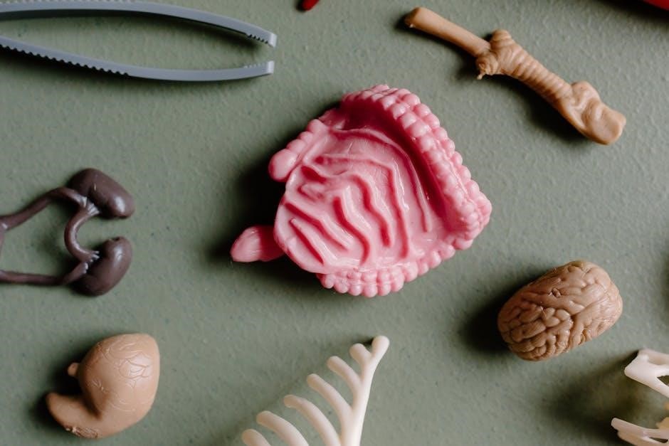
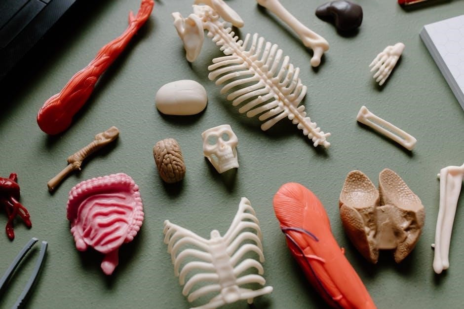
Visual Aids and Learning Tools
Visual aids like diagrams, 3D models, and illustrations simplify complex anatomical and physiological concepts, enhancing student understanding and engagement with the material․
10․1 Role of Diagrams and Illustrations
Diagrams and illustrations in anatomy and physiology lab manuals enhance understanding by visually representing complex structures and processes․ They make abstract concepts tangible, allowing students to explore details like cellular structures, muscle fibers, and organ systems․ These visuals support various learning styles and complement textual explanations, ensuring a comprehensive grasp of anatomical and physiological principles through clear, detailed representations․
10․2 Utilizing Online Resources for Enhanced Learning
Online resources, such as eLabs and digital platforms, complement anatomy and physiology lab manuals by offering interactive simulations, virtual dissections, and 3D models․ These tools provide immersive learning experiences, allowing students to explore complex anatomical structures and physiological processes in detail․ Accessible anytime, they support self-paced learning and reinforce concepts through engaging, technology-driven methods that enhance traditional lab-based education․